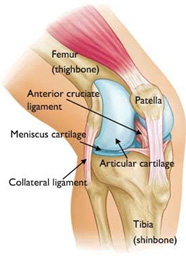
Anatomy of the Knee joint
The knee joint is the articulation between the thigh bone (Femur) and shin bone (Tibia). The main muscles that move the knee joint are the quadriceps, which is the front thigh muscle, and Hamstring (Back thigh) muscles. The Quadriceps muscle is attached to the tibial tubercle through patella tendon. The patella, or kneecap, is between the quadriceps tendon and the patella tendon.

The under surface of the kneecap articulates with a groove in the femur called the Trochlear. This arrangement of quadriceps tendon attachment, through the patella to tibia, increases the mechanical advantage of the quadriceps muscle.
There are four major ligaments in the knee joint, one on either side being the medial collateral ligament (inside ligament) and the lateral collateral ligament (outside ligament), and the cruciate ligaments which are two ligaments that cross over each other. The front cruciate ligament is the anterior cruciate ligament, and the back one, also the thickest ligament, is called posterior cruciate ligament. These ligaments, and associated muscles around the knee joint, provide the static and dynamic stability to the joint.
The articulating surfaces of the bones are covered with articular cartilage. The articular cartilage does not possess major repair capability and hence, when the articular cartilage is damaged it is not repaired by the body with similar articular cartilage. Usually articular cartilage damage is repaired by the body with fibrous cartilage which has inferior functional qualities to the normal articular cartilage. In addition to the articular cartilage two fibro-cartilaginous structures, meniscus, are attached to the rim of the tibia articular surfaces on either side of the knee.
The function of the Meniscus is to act as shock absorbing and position sensing structures.


